Optical Probe Displays Cancerous Tissue
 Jun-27-15
A new optical probe that causes cancerous tissue to appear red could help ensure surgeons remove only the unhealthy tissue during brain cancer surgeries.
Jun-27-15
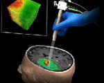
A new optical probe that causes cancerous tissue to appear red could help ensure surgeons remove only the unhealthy tissue during brain cancer surgeries.One of the trickiest parts of brain surgery is ensuring all of the tumor material is removed while leaving the neighboring tissue undamaged. In order to simplify the process, researchers at Johns Hopkins University have developed an optical coherence tomography (OCT) probe that can identify cancerous tissue in real-time.
Designed specifically for use during brain surgeries, the device was created based on the way brain cancer cells will scatter light. Using this knowledge, the team designed a computer program that recognizes the differences in the scattering of light and creates a 3D color map that shows the cancerous tissue in red, while the healthy tissue appears green.
More Info about this Invention:
[HOPKINSMEDICINE.ORG][STM.SCIENCEMAG.ORG]
Add Your Comment