3D Heart Models Simplify Medical Explanations
 Dec-30-13
By using a more realistic, skeletal 3D model, researchers have developed a way to create a 3D image of a baby’s heart structure, allowing the physician to share the information with laypeople.
Dec-30-13
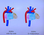
By using a more realistic, skeletal 3D model, researchers have developed a way to create a 3D image of a baby’s heart structure, allowing the physician to share the information with laypeople.Infants are not able to be placed in 3D scanners, so physicians usually rely on echo imaging and their own expertise to create a mental image of the baby’s heart. However, that mental image is not easy to share with other people—such as the baby’s parents. In order to present the information in the way an average person can understand, researchers developed a way to image the 3D information on a skeletal data structure, which is able to render blood vessels and organs and create a more realistic image.
The ultimate goal of the team is to create digital medical records on a variety of conditions, which could be retrieved as needed in order to allow the system to be used in clinical practice.
More Info about this Invention:
[DIGINFO.TV]
Add Comment
very nice
Posted by ayan king on December 31, 2013
fields are required.
Comments
very nice
Posted by ayan king on December 31, 2013
Add your Comment:
[LOGIN FIRST] if you're already a member.fields are required.
Show 1 Comment