Dye-Free Technique Images Cells in Real Time
 Jun-26-18
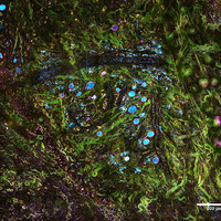
The SLAM microscopy technique can image living cells in real time, and could have significant applications in diagnosis and treatment.
Jun-26-18
The SLAM microscopy technique can image living cells in real time, and could have significant applications in diagnosis and treatment.The technique, from the team at the University of Illinois, eliminates the need for stains or dye, relying instead on pulses of light sent at different wavelengths. Called simultaneous label-free autofluorescence multi-harmonic microscopy (SLAM), the technology can image different tissues at the same time, offering an unprecedented level of cellular, including cellular communication.
More Info about this Invention:
[MEDGADGET.COM][NEWS.ILLINOIS.EDU]
Add Your Comment