Hyperspectral Microscope Sees Cancerous Tissue
 Oct-03-19
A hyperspectral surgical microscope able to identify cancer cells in real-time helps ensure the entire tumor has been removed.
Oct-03-19
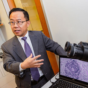
A hyperspectral surgical microscope able to identify cancer cells in real-time helps ensure the entire tumor has been removed.Developed by a team from the University of Texas at Dallas, the microscope analyzes tissue at several levels by imaging across most of the optical spectrum from UV to infrared. Machine learning is then applied to analyze the images for the signs of cancer. The results are nearly instant, reducing time and surgery costs—since standard frozen tissue takes up to 45 minutes to properly evaluate—while also reducing the amount of time a patient has to stay under anesthesia.
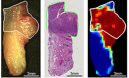
Image: Noninvasive hyperspectral imaging (right) compares to a current microscope method (center) in identifying cancer cells. The original tissue sample is on the left.
More Info about this Invention:
[MEDGADGET.COM][UTDALLAS.EDU]
Add Your Comment